Osseointegration Technology in 3D Printed Ti Implants
Osseointegration technology is redefining the performance benchmark for 3D printed titanium implants, where porous architecture, surface chemistry, and mechanical compatibility directly influence long-term fixation and patient recovery.
For technical evaluators, the question is no longer whether additive manufacturing can replicate trabecular bone.
The real issue is whether osseointegration technology can deliver reproducible pores, validated biocompatibility, and clinical evidence acceptable for high-risk implants.
Why Osseointegration Technology Needs a Checklist Approach
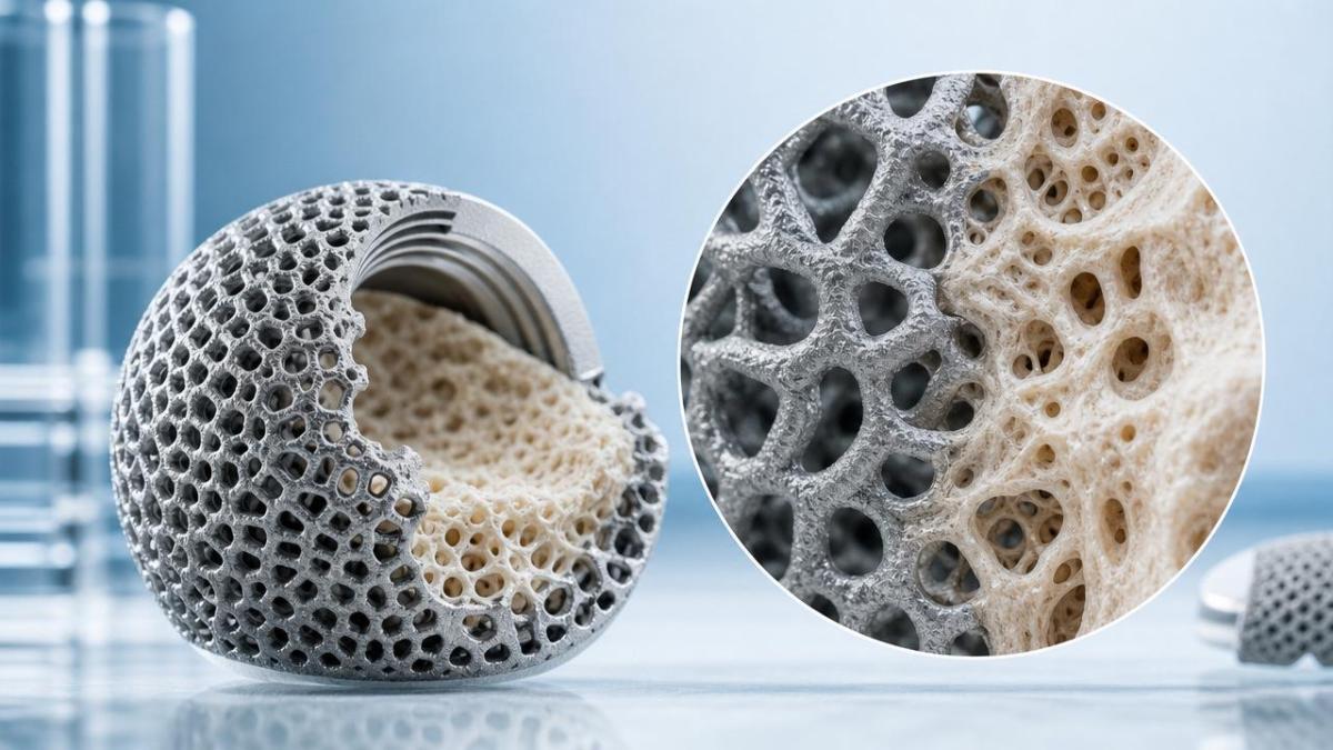
3D printed Ti implants succeed only when engineering intent, biological response, and regulatory documentation stay aligned.
A porous cup, spinal cage, or custom cranial plate may look advanced, yet fail if bone ingrowth remains superficial.
Osseointegration technology therefore requires structured evaluation across design, powder control, printing, cleaning, surface treatment, testing, and clinical follow-up.
This checklist format helps connect design parameters with patient outcomes.
It also supports technical dossiers, supplier qualification, and risk control under Class III medical device expectations.
Core Checklist for 3D Printed Ti Implant Osseointegration Technology
Use this checklist to assess whether osseointegration technology is engineered as a validated system, not only as a porous visual feature.
- Define the target anatomical load case before selecting pore size, because hip cups, cages, wedges, and custom implants transfer stress differently.
- Verify pore interconnectivity with micro-CT data, not only design files, to confirm continuous pathways for vascularized bone ingrowth.
- Control pore size distribution within a biologically meaningful range, commonly balancing osteoblast migration, capillary invasion, and fatigue strength.
- Match elastic modulus to surrounding bone where possible, reducing stress shielding while preserving structural safety under cyclic loading.
- Specify Ti6Al4V ELI or equivalent implant-grade titanium with traceable chemistry, powder history, oxygen content, and reuse limits.
- Validate powder removal from internal lattice zones, because retained particles may affect wear debris, inflammation, and long-term biological safety.
- Inspect roughness at macro, micro, and nano levels, since osseointegration technology depends on multi-scale cell attachment signals.
- Confirm surface chemistry after blasting, acid etching, passivation, or coating, especially oxide stability and contamination control.
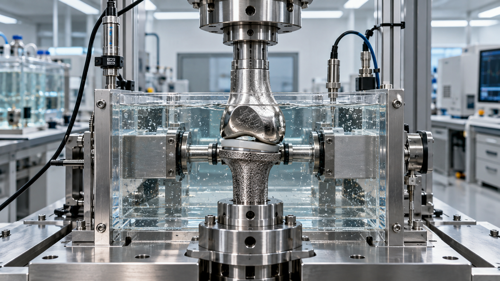
- Run fatigue testing on final geometry, because lattice structures may introduce local stress concentrations invisible in static compression tests.
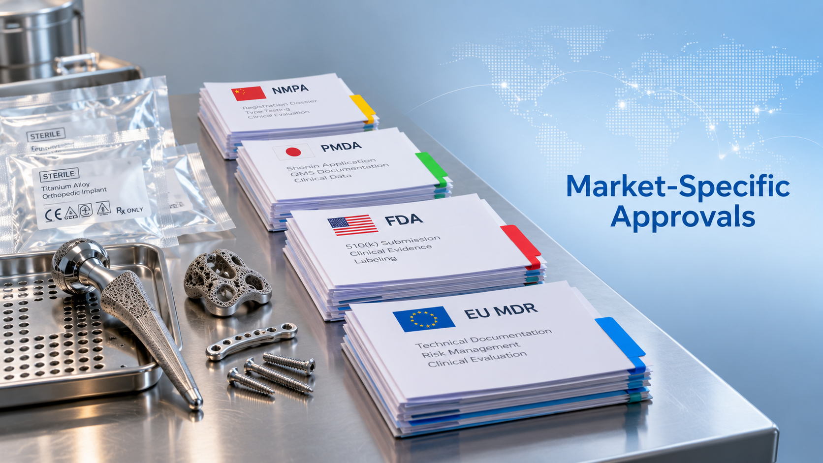
- Link every design claim to bench, biological, and clinical evidence, ensuring osseointegration technology can withstand regulatory review.
Design Factors That Decide Bone Ingrowth Performance
Porous Architecture
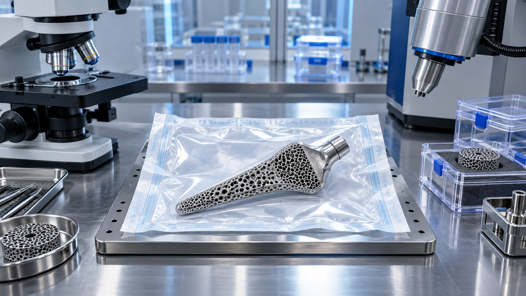
Porous architecture is the visible face of osseointegration technology, but geometry alone does not guarantee fixation.
Pore size, strut thickness, permeability, tortuosity, and surface area must function together.
Highly porous structures may support early vascular penetration, yet excessive porosity can reduce mechanical endurance.
A balanced lattice should invite bone while surviving implantation forces, postoperative loading, and millions of gait cycles.
Surface Topography
Surface topography determines how proteins adsorb, cells attach, and osteogenic signaling begins.
In practical osseointegration technology, roughness should be intentionally created and consistently measured.
Uncontrolled partially melted particles can look rough, but they are not equivalent to validated micro-texturing.
The key is reproducible topography with stable titanium oxide and minimal loose debris.
Mechanical Compatibility
Mechanical compatibility is central to osseointegration technology because bone adapts to strain.
A very stiff implant may transfer insufficient load to adjacent bone.
A weak implant may deform, migrate, or fracture before mature bone bridges the interface.
Design review should therefore include modulus, yield strength, fatigue limit, and fixation stability.
Material and Manufacturing Controls for Reliable Osseointegration Technology
Additive manufacturing turns powder, energy, atmosphere, and scan strategy into implant structure.
Each variable can influence osseointegration technology by changing roughness, porosity, residual stress, or contamination risk.
- Audit powder lifecycle, including supplier qualification, particle size distribution, morphology, reuse cycles, and segregation during handling.
- Monitor build atmosphere and oxygen exposure, since titanium reactivity can alter ductility, oxide behavior, and fatigue performance.
- Control laser parameters and scan paths, preventing lack-of-fusion defects, keyhole porosity, and inconsistent lattice strut formation.
- Apply validated heat treatment to relieve residual stress without degrading dimensional accuracy or microstructural stability.
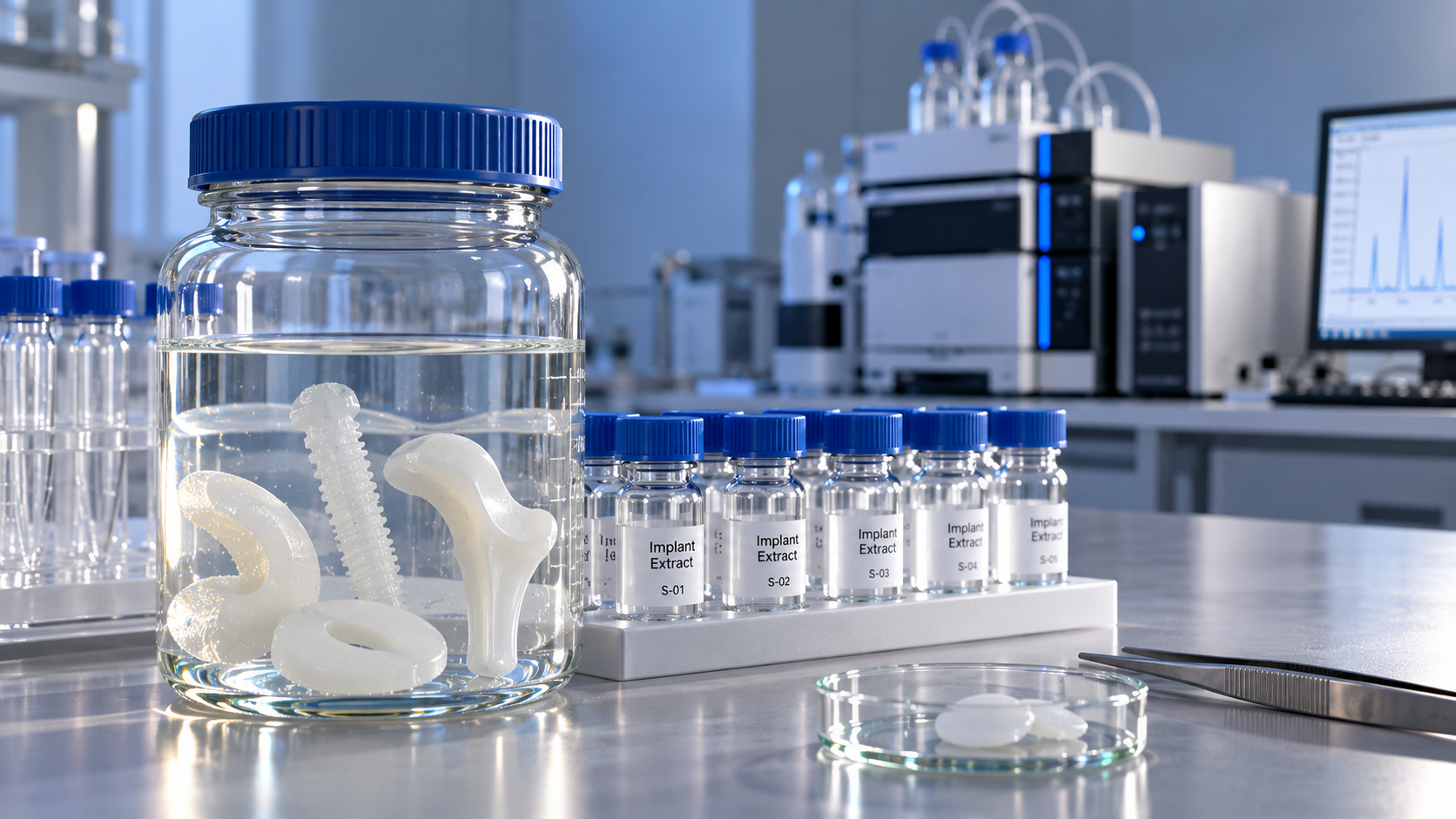
- Document cleaning effectiveness for porous implants, using extraction studies, particle analysis, and residue evaluation.
- Maintain lot-level traceability from powder batch to finished implant, supporting recalls, audits, and post-market surveillance.
Cleaning deserves special attention in osseointegration technology.
Deep lattices can trap unmelted powder, blasting media, machining fluid, or chemical residues.
Visual inspection is insufficient when internal porous zones cannot be directly observed.
Biological Safety and Regulatory Evidence
Osseointegration technology must pass beyond attractive imaging and demonstrate biological safety.
For titanium implants, ISO 10993 evaluation usually addresses cytotoxicity, sensitization, irritation, systemic toxicity, genotoxicity, implantation, and chemical characterization.
The biological evaluation should reflect the final sterilized device, not an idealized coupon.
Coupons help screening, but final device testing better captures lattice complexity, cleaning limits, and surface modification effects.
Clinical evidence is equally important for osseointegration technology in high-risk orthopedic implants.
Radiographic bone ingrowth, migration analysis, revision rates, pain scores, and functional recovery can support performance claims.
Regulatory submissions should connect preclinical mechanisms with clinical endpoints.
A strong file explains why the selected porous structure should improve fixation without introducing new unacceptable risks.
Application Notes Across Implant Scenarios
Hip and Knee Reconstruction
In acetabular cups and revision components, osseointegration technology focuses on initial press-fit stability and long-term bone ingrowth.
The porous layer should resist shear, preserve hemispherical accuracy, and avoid excessive stiffness at the bone interface.
For knee cones, sleeves, and augments, defect filling and load transfer become decisive.
The lattice must support metaphyseal fixation while allowing surgeons to achieve stable intraoperative positioning.
Spinal Interbody Cages
Spinal cages use osseointegration technology to encourage endplate fusion and reduce subsidence risk.
Porosity must balance bone graft integration, imaging visibility, compression strength, and contact area.
Surface roughness may enhance stability, but aggressive textures can damage endplates during insertion.
A cage design should consider surgical approach, lordotic angle, graft window, and radiographic assessment.
Patient-Specific Implants
Patient-specific Ti implants extend osseointegration technology into complex trauma, oncology, and craniofacial reconstruction.
Customization improves anatomical fit, but every unique geometry complicates validation and documentation.
Parametric design rules, locked process windows, and case-based verification help maintain consistency.
The implant should be personalized without becoming biologically or mechanically unpredictable.
Commonly Overlooked Risks in Osseointegration Technology
Overclaiming trabecular similarity: A lattice resembling cancellous bone does not prove equivalent biological performance.
Claims should be supported by measurable pore geometry, mechanical tests, animal data when needed, and clinical performance signals.
Ignoring interface micromotion: Bone ingrowth may fail when early micromotion exceeds tolerable limits.
Osseointegration technology should be evaluated with fixation method, surgical technique, and postoperative loading in mind.
Underestimating sterilization effects: Sterilization can influence packaging residues, surface chemistry, and material aging.
Testing should reflect the final sterilized, packaged, and shelf-aged device whenever justified by risk analysis.
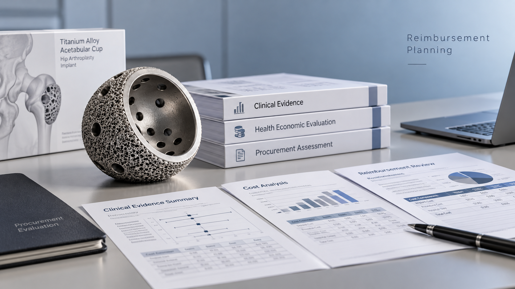
Separating VBP pressure from quality: Cost control can encourage simplified processes, but porous Ti implants require disciplined validation.
Price competition should not reduce cleaning controls, fatigue testing, or biological evaluation depth.
Practical Execution Advice
- Create a design history map linking every osseointegration technology feature to a biological or mechanical rationale.
- Use micro-CT as a routine verification tool for porous zones, especially after parameter changes or supplier transfer.
- Set acceptance criteria for roughness, porosity, residual powder, and fatigue strength before production scaling begins.
- Build a biological safety plan around the final device, final cleaning process, and final sterilization method.
- Collect post-market data on loosening, migration, infection, revision, and radiographic integration to refine future designs.
Execution should remain cross-functional.
Design engineering, materials science, toxicology, clinical science, manufacturing, and regulatory strategy must work from the same evidence chain.
This integrated approach reflects the future of osseointegration technology in premium orthopedic reconstruction.
Summary and Action Guide
3D printed Ti implants have moved osseointegration technology from coating strategy to full structural design philosophy.
Successful bone ingrowth depends on reproducible porosity, clean surfaces, compatible mechanics, and evidence that survives clinical and regulatory scrutiny.
The next step is to review one implant platform against the checklist above.
Start with pore verification, powder traceability, cleaning validation, fatigue evidence, and ISO 10993 biological evaluation.
Then connect these findings to clinical endpoints such as fixation stability, bone ingrowth, revision reduction, and patient function.
When osseointegration technology is managed as a validated system, 3D printed titanium implants can better reconstruct mobility, durability, and long-term patient confidence.
Get weekly intelligence in your inbox.
No noise. No sponsored content. Pure intelligence.
News Recommendations
 Oct 26, 2026Can a single approval cover life science product sales worldwide?Can a single approval cover life science product sales worldwide?
 Oct 25, 2026When is sensitization testing needed for medical device biocompatibility?When is sensitization testing needed for medical device biocompatibility?
 Oct 24, 2026How is biocompatibility tested for implantable polymer biomaterials?How is biocompatibility tested for implantable polymer biomaterials?
 Oct 23, 2026How to build reimbursement into an implant commercialization strategyHow to build reimbursement into an implant commercialization strategy
 Oct 22, 2026What clinical evidence do implants need for CE marking under EU MDR?What clinical evidence do implants need for CE marking under EU MDR?
 Oct 21, 2026How to choose disposable laparoscopic stapler reloads for thick tissueHow to choose disposable laparoscopic stapler reloads for thick tissue
 Oct 20, 2026How to assess medical polymer biocompatibility under ISO 10993How to assess medical polymer biocompatibility under ISO 10993
 Oct 19, 2026How do interventional device categories differ in clinical use?How do interventional device categories differ in clinical use?
 Oct 18, 2026When to choose alginate dressings for heavily exuding chronic woundsWhen to choose alginate dressings for heavily exuding chronic wounds
 Oct 17, 2026How is biocompatibility tested for custom medical-grade PEEK implants?How is biocompatibility tested for custom medical-grade PEEK implants?
 Oct 16, 2026How to check a wound healing materials supplier’s biocompatibility dataHow to check a wound healing materials supplier’s biocompatibility data
 Oct 15, 2026How to verify the FDA clearance status of silver wound dressingsHow to verify the FDA clearance status of silver wound dressings
 Oct 14, 2026How to register orthopedic implants and instruments in Latin AmericaHow to register orthopedic implants and instruments in Latin America
 Oct 13, 2026How to use a device market intelligence database to benchmark VBP pricesHow to use a device market intelligence database to benchmark VBP prices
 Oct 12, 2026When should stainless steel orthopedic instruments be replaced?When should stainless steel orthopedic instruments be replaced?
 Oct 11, 2026When are alginate dressings rope the right choice for cavity wounds?When are alginate dressings rope the right choice for cavity wounds?
 Oct 10, 2026When smart minimally invasive technology improves catheter accessWhen smart minimally invasive technology improves catheter access
 Oct 08, 2026Clinical evaluation requirements for cardiovascular implant devices in EuropeClinical evaluation requirements for cardiovascular implant devices in Europe
 Oct 07, 2026What to verify before appointing an artificial joint implants distributorWhat to verify before appointing an artificial joint implants distributor
 Oct 06, 2026How ISO 10993 guides biocompatibility testing for medical materialsHow ISO 10993 guides biocompatibility testing for medical materials
 Oct 05, 2026How to verify an orthopedic implants and instruments exporterHow to verify an orthopedic implants and instruments exporter
 Oct 04, 2026What drives surgical stapling devices price differences in hospital tenders?What drives surgical stapling devices price differences in hospital tenders?
 Oct 03, 2026How to assess a central venous catheter manufacturer's infection-control evidenceHow to assess a central venous catheter manufacturer's infection-control evidence
 Oct 02, 2026How to assess an advanced wound care factory’s quality systemHow to assess an advanced wound care factory’s quality system
 Oct 01, 2026What is driving global medical consumables demand in emerging markets?What is driving global medical consumables demand in emerging markets?
 Sep 30, 2026Implant Material Compliance Testing: Evidence Needed for Regulatory SubmissionImplant Material Compliance Testing: Evidence Needed for Regulatory Submission
 Sep 30, 2026From Corrosion to Fatigue: What Implant Materials Testing Should MeasureFrom Corrosion to Fatigue: What Implant Materials Testing Should Measure